Preview
Media Type
Image
Description
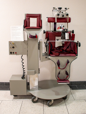
The early brain imaging (x-ray) techniques beginning in 1919 involved the practice of injecting air into the spinal canal (pneumoencephalography) and rolling the patient about in a specially devised chair which is shown here. These patients were often uncomfortable, developed severe headaches, and became nauseated. Perhaps the greatest contribution of CT and MRI imaging, in the mid 1970s, was to make pneumoencephalography obsolete. This chair was donated by Lee Memorial Hospital.
Keywords
Museum of Medical History, special collections, image gallery