

Preview
Description
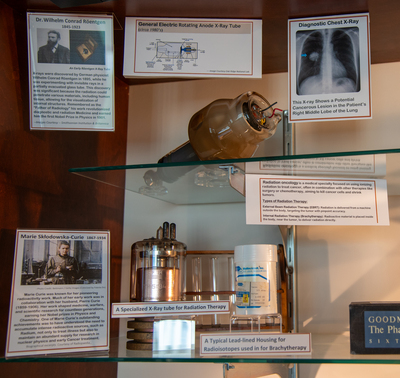
The left upper corner of the Oncology Display provides a tribute to Dr. Wilhelm Conrad Röntgen, the first recipient of a Nobel Prize in Physics in 1901. His seminal work both in the discovery of how to produce x-rays and the diagnostic and early therapeutic consequences for this technology provided a revolution in both the diagnosis and early treatment of cancer. This technology is essential for early diagnosis and guides treatment for a myriad of human disease and injury. A diagram as well as an actual functional rotating anode type x-ray tube that resulted from Röntgen’s early work is displayed on the top shelf. An example of a chest x-ray image, also known as a roentgenograph, illustrates this technology for early detection of a suspicious lesion or shadow in the human right lung is also displayed to the left.
The left side of the second shelf provides a tribute to Marie Curie who received two Nobel Prizes for Physics and Chemistry. Her discovery and later work with radium and other radioactive substances paved the way for radiation therapy. Artifacts to the left of her image are designed to further illustrate Marie Curie’s contributions; they include the wooden rack of glass test tubes from a chemistry lab, a specialized tube for generating x-rays for radiation therapy, and a lead-lined container for transport of radioisotopes. Above these objects is a storyboard with a definition of Radiation Oncology and its two branches.
Media Type
JPG
Keywords
Museum of Medical History, special collections, image gallery, Gulf Coast Medical Center